


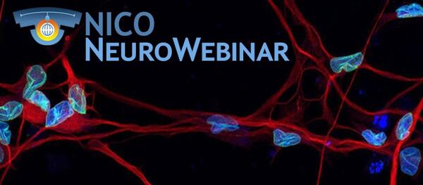
NeuroWebinar & Seminar
1 appointment per week, on Friday at 2.00 pm
**Hybrid seminar: both in presence (max 25 people in Seminar room) and on webex
Wednesday 30/7/2025 h. 11.00 am
- Hybrid seminar
Prof. Meltem Bahcelioglu, Head of Anatomy & Neuroscience Departments, Gazi University, Faculty of Medicine, Deputy Director and Executive Board Member, Neuroscience and Neurotechnology Center of Excellence (NÖROM), Ankara, TÜRKİYE
NÖROM—Neuroscience and Neurotechnology Center of Excellence: First Joint Research Center in Türkiye
NÖROM, the Neuroscience and Neurotechnology Center of Excellence, is Türkiye’s pioneering hub for brain research, founded on a unique partnership between three of the nation's leading institutions: Ankara University, Gazi University, and Middle East Technical University (METU). Established as the first joint research center of its kind by the Turkish Higher Education Council (YOK), NOROM brings together the fields of medicine, fundamental sciences, and engineering. Its core mission is to create a sustainable neuroscience ecosystem that produces innovative knowledge and technology for the benefit of humanity.
NOROM's more than 67 academic staff members and researchers leading groups focused on high-impact areas such as deep learning and computational neuroscience, migraine and pain, cognitive neuroimaging, the brain-gut axis, neurodevolepmantel disorders (Autism spectrum disorders, ADHD), balance and walking, and neuro-rehabilitation. This collaborative environment has proven highly productive, with researchers securing significant funding and producing 129 articles in international journals (H-index: 12), while also winning numerous awards for their innovative work.
NOROM welcomes international partnerships with universities, research institutions, and industry to pioneer the future of neuroscience.
Host: Corrado Calì| webex link
2025 - ARCHIVE
Friday 18/7/2025 h. 2.00 pm - Webinar
Salman Zubedat, Spatial Perception and Memory Lab, Department of Neuroscience, The Rappaport Faculty of Medicine, Technion-Israel Institute of Technology, Israel.
Large increase in dentate gyrus activity accompanies aggression in a resident-intruder task
Territoriality entails behaviors in which an individual claims and defends a spatially confined area from conspecifics. Our goal was to explore its neural foundations. We hypothesized that territoriality is supported by neural circuits linking memory and spatial representation, with the hippocampus playing a central role. To investigate this, we conducted calcium imaging of the dentate gyrus in the dorsal hippocampus during a resident-intruder paradigm. In this setup, a male ICR mouse expressing calcium indicators in the dentate gyrus was housed with a sterile female for 21 days to establish territoriality. Starting on day 21, the resident mouse was exposed to an intruder for 10-minute sessions over several days.
Initially, no notable changes were observed in behavior or neural activity. However, after 3-5 days, the number of active cells in the dentate gyrus increased by 3 to over 10 times the original level, coinciding with a shift from friendly to aggressive behavior. Interestingly, the aggressive behavior persisted even after the activity of the dentate gyrus subsided.
These findings suggest that significant plastic changes occur in the dentate gyrus following the introduction of an intruder, highlighting its capacity to dramatically alter its activity in response to pivotal experiences.
Host: Serena Bovetti
Friday 11/7/2025 h. 2.00 pm - Hybrid seminar
Dr. Michael Tsoory, Weizmann Institute of Science, Israel
Detecting Sciatic nerve injury (SNI) induced motor impairments and recovery using the a home-cage monitoring system; a core facility unit user’s perspective
The talk focuses on experiments conducted using the DVC® system in our facility. It describes the validation processes and the development of a “data pipeline”.
It will detail experiments aimed to assess the DVC® system’s capacity to detect changes in home cage activity that stem from induced motor impairments and recovering from them.
Laboratory rodent models of nerve injury rely heavily on repeated assessments of motor functions away from the home–cage, where the laboratory animals are often forced to walk to allow assessments of gait and stride, for example. Such assessments lead to substantial disturbance of the animals’ routine and cause them some discomfort that might mask the experimental manipulation effects. In addition, these evaluations are labor-intensive and require time-consuming post-hoc analyses.
Therefore, the described study sought a home-cage-based alternative and in a series of experiments assessed the DVC® system’s capacity to detect sciatic nerve injury-induced motor impairments and recovery dynamics as reflected by changes in voluntary, spontaneous, activity in the home cage.
The home – cage activity DVC® indices data indicate dynamics of recovery processes, reflected by “return to baseline levels”, that correspond to those indicated by stride and gait analyses. Additional discussion will address the advantages and challenges of monitoring home cage activity from a core facility user’s perspective. The lecture discusses in a critical manner issues of validity, reliability, scientific rigor and animal well fare.
Host: Letizia Marvaldi
Friday 4/7/2025 h. 2.00 pm - Hybrid seminar
PROGRESS REPORT
Stefano Zucca (Adult Neurogenesis Group)
Multisensory Integration of Social Cues in the ventromedial Prefrontal Cortex
The ability to attract and reproduce with a suitable mate is a key driver of evolution. Mate selection depends on social interaction, and animals have evolved diverse behavioural strategies that rely on multimodal communication. Despite the importance of multisensory cues in courtship, the neural circuits responsible for integrating these signals remain poorly understood. In rodents, males use a combination of olfactory (e.g., pheromones) and acoustic (ultrasonic vocalizations, USVs) cues to court females. Recent studies show that female mice prefer USVs from unfamiliar males only when paired with sexual odors, suggesting cross-modal integration. However, where and how this sensory information converges in the brain remains unclear. We first confirmed through behavioural preference tests that female mice favor multimodal cues, but only within a social context. To explore the brain’s response to unimodal and multimodal stimuli, we conducted whole-brain analysis of immediate-early gene (IEG) expression, using tissue clearing and light-sheet microscopy. Our results revealed that sexual odors predominantly drive brain activation, with the ventromedial prefrontal cortex (vmPFC) specifically responding to multisensory social cues. To further investigate vmPFC involvement, we used miniscope recording to perform one-photon functional imaging in awake animals. We found that vmPFC neurons respond to both unimodal and multimodal cues, with a distinct subset selectively activated by combined sensory inputs. By performing chemogenetic manipulation of vmPFC neurons, we are now investigating their causal role in shaping female preference for multisensory social stimuli. Together, these findings highlight the vmPFC as a key hub for integrating social cues from different modalities, suggesting its potential role in shaping mate preference.
Friday 20/6/2025 h. 2.00 pm - Hybrid seminar
Daniela Ferrari, University of Milano-Bicocca - Department of Biotechnology and Biosciences
Investigational cell-based treatments using neural stem cells for Amyotrophic Lateral Sclerosis
Cell-based strategies continue to be among the most promising avenues for developing effective experimental treatments for disorders of the central nervous system. Due to their remarkable functional adaptability, stem cells offer a wide range of therapeutic possibilities, including cell replacement, immune system modulation, anti-inflammatory actions, trophic support and toxicitybluntingeffects. In this context, I will provide an overview of our work and highlight key findings from both preclinical and clinical studies that have explored the therapeutic potential of neural stem cells, with a particular focus on their application in treating Amyotrophic Lateral Sclerosis (ALS).
Host: Marina Boido
SEMINAR CYCLE of the PhD in Neuroscience of Turin
Wednesday 18/6/2025 h. 11.00 am - Hybrid seminar
Cornelius Gross, Epigenetics & Neurobiology Unit, European Molecular Biology Laboratory, EMBL Rome
How does context and experience shape social fear circuits?
Exposure to predators or predator-like stimuli elicit powerful negative emotions and uncontrollable escape responses across animal species. Over the last decade we have dissected the brain circuits that mediate such innate threat responses in mice in order to learn more about human fear. We have identified the brain pathways that mediate innate responses to predators and shown that these are independent from those that mediate responses to social threats. We have recorded the responses of individual neurons while animals initiate escape from threat and have identified a local microcircuit that supports the escape decision.
More recently we have turned to studying the neural circuits supporting avoidance of social threats.
We have found that social hierarchy and social context can modulate the threshold for escape from social threats, showing that innate emotional behavior responses can be reshaped by experience. We are currently exploring how territoriality impacts social fear and aggression, and how our neurocircuit findings in the mouse may be relevant for human social behavior.
My talk will be preceded by a short overview of EMBL.
Host: Serena Bovetti
Friday 13/6/2025 h. 2.00 pm - Webinar
Paul Weiringa, Maastricht University, MERLN - Institute for Technology-Inspired Regenerative Medicine
The Peripheral Nervous System in Tissue Engineering: from Scaffold Design to Functional In Vitro Models
The peripheral nervous system (PNS) plays a critical role in tissue function, homeostasis and repair, making it a compelling yet underappreciated target for tissue engineering and regenerative medicine. This lecture outlines the fundamentals of peripheral nerve biology and its broader relevance beyond nerve repair, including its influence on the regeneration and pathology of other tissues. Emphasis is placed on biofabrication strategies for engineering PNS-relevant environments, including electrospinning and melt electrowriting (MEW) technologies for producing fibrous scaffolds that mimic the extracellular matrix. Challenges and advancements in generating controlled architectures, such as microchannel networks that replicate native tissue organization, will also be discussed. Highlighting work from our lab, the lecture will showcase the development of complex 3D in vitro models aimed at recreating functional tissue-nerve interfaces in different contexts. These integrated approaches aim to advance understanding of tissue innervation in health and disease while advancing the boundaries of biofabrication.
Host: Stefania Raimondo | webex link
Friday 9/5/2025 h. 2.00 pm - Webinar
António J. Salgado, Life and Health Sciences Research Institute (ICVS), School of Medicine, University of Minho, Braga, Portugal; ICVS/3Bs PT Associated Lab, Braga/Guimarães, Portugal
Stem Cells Secretome in Central Nervous System Regenerative Medicine: Insights from Pre-Clinical Models of Injury and degeneration
The low regeneration potential of the central nervous system (CNS) represents a challenge for the development of new therapeutic strategies. Mesenchymal stem cells (MSCs) have been proposed as a possible therapeutic tool for CNS disorders, namely due to the beneficial actions of their secretome. Indeed, the latter possesses a broad range of neuroregulatory factors that promote an increase in neurogenesis, inhibition of apoptosis/glial scar, immunomodulation, angiogenesis, neuronal and glial cell survival, as well as relevant neuroprotective actions into different pathophysiological contexts. Considering their protective action in lesioned sites, MSCs, and their secretome, might also improve the integration of local progenitor cells in neuroregeneration processes. In this sense their use could represent an important vehicle for the establishment of future CNS regenerative therapies. Previously we have shown that the administration of MSCs secretome in pre-clinical models of spinal cord injury (SCI) and Parkinson’s Disease (PD) led to important motor and histological improvements. In order to further improve this, in the present talk we will discuss and present the outcomes of the combinatory use of MSCs secretome and biodegrable biomaterials, particularly self-assembling peptides, lypossomes and hydrogels in in vitro and in vivo models of SCI and PD.
Host: Stefania Raimondo | webex link
Tuesday 6/5/2025 h. 11.00 am - Hybrid seminar
Department of Neuroscience Rita Levi Montalcini, Unito
Main Hall (Aula Magna Neurologia), 1 piano, via Cherasco 15, Torino
Serenella Tolomeo, Institute of High Performance Computing, A*STAR
Neuroimaging in Psychiatry
Dr Tolomeo will present her research in the field of neuroimaging, cognitive neuroscience and psychiatry. The implications for prevention, intervention and future directions.
Host: Alessandro Vercelli
Tuesday 1/4/2025 h. 4.30 pm - Hybrid seminar
Javier DeFelipe, Laboratorio Cajal de Circuitos Corticales (CTB), Universidad Politécnica de Madrid and Instituto Cajal (CSIC), Madrid
Brain connectomics: exploring the connectome and synaptome
The principal goal in neuroanatomy is to define the detailed structural design of the nervous system. This challenge is one of the first steps towards understanding how neural circuits contribute to the functional organization of the nervous system, both in health and disease. The main difficulties involve unraveling the extraordinary complexity of the nervous system and to define how information flows through this finely organized synaptic network. Over the years, neuroanatomy has evolved considerably thanks to the use of classical techniques and the introduction of new procedures. The term “connectome” has recently been proposed to refer to the highly organized connection matrix of the human brain, in analogy to the human genome. However, defining how information flows through such a complex system represents so difficult a task that it would seem unlikely it could be achieved in the near future, or, for the most pessimistic, perhaps never. Circuit diagrams of the nervous system can be considered at different levels, although they are surely impossible to complete at the synaptic level. Even for a small mammal like the mouse it is impossible to fully reconstruct the brain at this level (we would need over 1.4 x 10⁹ sections to fully reconstruct just one mm3 of tissue). Therefore, complete reconstructions of a small region of the mammalian brain are feasible, while structures like the cerebral cortex cannot be fully reconstructed. Despite the technical difficulties, by adopting appropriate strategies with the tools now available coupled with the development of huge international projects, it should be possible to make spectacular advances in unraveling brain organization, even in humans. Indeed, advances in our capacity to marry macro- and microscopic data may help establish a realistic statistical model that could describe connectivity at the ultrastructural level, the “synaptome”, giving us cause for optimism.
Host: Alessandro Vercelli
Friday 28/3/2025 h. 2.00 pm - Webinar
Maria Ludovica Sforza, Laboratory of Neural Epigenomics, Institute for Medical Physics and Micro-tissue Engineering, Friedrich-Alexander-Universität Erlangen-Nürnberg (FAU), Erlangen, Germany; Laboratory of Nuclear Architecture in Neural Plasticity and Aging, German Center for Neurodegenerative Diseases (DZNE), Dresden, Germany
Investigating the role of Nup153 in neuronal responsiveness and its link to cognition
Neurons respond dynamically to environmental stimuli through the expression of the so-called immediate early genes (IEGs). Accurate spatio-temporal activation and repression of IEGs is vital for encoding specificity of incoming information and critical for proper neuronal plasticity, impacting on learning and memory. Nuclear pore complex proteins (Nups) play critical roles in cell type-specific gene regulation by orchestrating lineage specification and maintenance of cellular state. However, their roles in the adult brain remain largely elusive, especially in post-mitotic neurons. We hypothesize that Nups play key roles in neuronal plasticity and cognition through gene regulation. To address the importance of Nups for cognition and memory, we conducted behaviour, histological and electrophysiological assays on Nup153 heterozygous mice, one of key Nups in gene regulation.
Host: Francesca Montarolo
Friday 21/2/2025 h. 2.00 pm - Hybrid seminar
Katja Reinhard, Flexibility in Circuits & Behaviour Lab, SISSA - Trieste, Italy
The neural basis of context-dependent behaviour adaptation
Avoiding danger is one of the most essential and conserved sets of behaviours, observed in most species from crabs to primates. To optimize an animal’s survival, avoidance responses need to be fast and reliable, but also flexible and adaptable to the current context. However, how this flexibility in behavioural output is implemented in the brain is largely unknown. Furthermore, it is unclear how similar the precise circuits and mechanisms underlying a conserved behaviour are across contexts and species.
The goal of my lab is to identify how information about the environment and state can adapt behavioural decision making. We approach this by using a highly standardized behaviour assay where we compare innate reactions and neural circuit activity while changing selected contextual elements. By probing the same behaviours and circuits under different circumstances, we aim to identify principles of behavioural flexibility that are conserved across contexts and species as well as to reveal elements that are most likely to change. During this seminar, I will focus on the brain circuit architecture that allows for contextual information to shape innate behaviours and talk about how habitat differences have led to permanent changes in innate behaviours and specific underlying circuit nodes.
Host: Stefano Zucca
Friday 7/2/2025 h. 3.00 pm
- Webinar
Roberta Piovesana, Department of Neurosciences, Université de Montréal - Groupe de Recherche sur la Signalisation Neurale et la Circuiterie, Université de Montréal, Canada
Novel role of endocannabinoid in neuromuscular junction structure and function
The peripheral nervous system has a remarkable ability to regenerate. Following nerve injury, different events happen at the axonal and neuromuscular junction (NMJ) level with the goal to generate an innervation-permissive environment. Several injury signals emerging from the injured axons and axon terminals have been identified to trigger the injury responses of Perisynaptic Schwann cells (PSCs), glial cells at the NMJ, essential for NMJ maintenance and repair. Although these NMJ-forming processes can be repeated, severe NMJ diseases can occur if a single step of NMJ formation is compromised. Thus, a better understanding of NMJ formation and maturation will help to understand both the regenerative processes that occur after nerve injury and how altered maturation leads to major defects.
Cannabinoids are frequently used in the treatment of neuropathic pain. However, despite evidence for their roles in the regulation of axonal guidance and synapse formation, their possible contribution in response to peripheral nerve injury remains unclear. In this webinar, we will explore the latest discovery on the glial Cannabinoid type 1 receptors at the NMJsfollowing nerve injury. Our results highlight a novel role of endocannabinoids in motor recovery, opening a possible therapeutic strategy for facilitating nerve repair or to address inadequate NMJ maintenance observed in motor neuron-related neurodegenerative diseases.
Host: Roberta Schellino
SEMINAR CYCLE of the PhD in Neuroscience of Turin
Thursday 30/1/2025 h. 2.00 pm - Hybrid seminar
Paolo Malatesta, University of Genoa and IRCCS Policlinico San Martino, Genoa
Tracing Clonal Evolution and Immune Evasion in Glioblastoma Progression
Human Glioblastoma remains a significant challenge in oncology research, with its early progression remaining notably elusive. In a recent study, we traced the clonal dynamics of glioblastoma evolution by co-introducing PDGFB and genetic barcodes into mouse brains, observing a sustained loss of clones during the transition to a malignant state—an effect tied to shifts in c-Myc expression levels and their downstream targets. We then extended our investigation of glioblastoma evolution by transplanting multiclonal, early- stage glioma cells into multiple immunodeficient NOD-SCID mice.
This approach allowed us to follow clonal behavior across serial transplants, revealing the acquisition of immune-evasive capabilities by early-stage glioma clones, which later proved able to initiate tertiary tumors in immunocompetent hosts. By combining barcode sequencing and single-cell RNA sequencing of early-stage gliomas with bulk RNA sequencing of secondary and tertiary tumors, we examined both the clonal and transcriptomic makeup of these lesions.
Among the numerous clones present in the primary tumors, only a fraction persisted through subsequent passages. Moreover, the clonal configuration of secondary tumors originating from the same primary glioma demonstrated partial overlap, hinting at a degree of predetermination in the acquisition of immune-evasive features. Our intra- and inter-clonal transcriptomic analyses across different tumor stages illuminate how novel functional traits may arise in gliomas.
Ongoing exploration of these data will reveal whether such traits stem from the expansion of clones already harboring them, or if they emerge through functional shifts within the clones themselves. Overall, our findings reinforce the significance of clonal competition, underscoring the central role of immune-system interactions in shaping these competitive dynamics.
Host: Federico Luzzati
SEMINAR CYCLE of the PhD in Neuroscience of Turin
Friday 31/1/2025 h. 2.00 pm - Hybrid seminar
Benedikt Berninger, King’s College London, UK and University Medical Center Mainz, Germany
Engineering neurogenesis in the postnatal cerebral cortex by lineage reprogramming
Lineage reprogramming of glia into neurons emerges as an experimental strategy to regenerate neurons lost to disease. We explore the possibility of using proneural transcription factors and mutant variants thereof to convert cortical glia (astrocytes and oligodendrocyte progenitor cells) into induced neurons with subtype specific properties. For example, we could show that forced expression of a phospho-deficient form of achaete-scute complex like 1 (Ascl1), referred to as Ascl1SA6, together with the cell death regulator Bcl2 can promote the conversion of early postnatal astrocytes into induced neurons that feature hallmarks of fast-spiking parvalbumin-expression interneurons (Marichal et al., 2024).
Here, I will discuss how much we have learned about the transcriptional programmes underlying the conversion process, how much induced neurons resemble/differ from their endogenous counterparts, how we aim at closing the gap between induced and endogenous neurons, and to which induced neurons succeed to integrate into functional cortical circuits.
Host: Federico Luzzati
Friday 17/1/2025 h. 2.00 pm - Hybrid seminar
Salvatore Adinolfi, Dipartimento di Scienza e Tecnologia del Farmaco, Università di Torino
Frataxin: a biochemistry tale from molecular to pathophysiology of neurodegeneration
Frataxin, a highly conserved protein found in prokaryotes and eukaryotes, is required for efficient regulation of cellular iron homeostasis. Humans with a frataxin deficiency have the cardio- and neurodegenerative disorder Friedreich’s ataxia, commonly resulting from a GAA trinucleotide repeat expansion in the frataxin gene.
I will revisit the most significant milestones obtained with biochemical and biophysical approaches that have led us to our current understanding of frataxin and its functions. While frataxin function remains a point of controversy, a clearer picture emerges suggesting that frataxin is an essential element of the crucial Iron-sulfur cluster biogenesis pathway.
Host: Ferdinando Di Cunto
2024
Friday 20/12/2024 h. 10.00 am
- Hybrid seminar
Gabriele Chelini, CNR Pisa
Focal cluster of peri-synaptic matrix contribute to activity-dependent plasticity and memory in mice
Recent findings show that effective integration of novel information in the brain requires coordinated processes of homo- and heterosynaptic plasticity. In this work, we hypothesize that activity-dependent remodeling of the peri-synaptic extracellular matrix (ECM) contributes to these processes. We show that clusters of the peri-synaptic ECM, recognized by CS56 antibody, emerge in response to sensory stimuli, showing temporal and spatial coincidence with dendritic spine plasticity. Using CS56 co-immunoprecipitation of synaptosomal proteins, we identify several molecules involved in Ca2+ signaling, vesicle cycling, and AMPA-receptor exocytosis, thus suggesting a role in long-term potentiation (LTP). Finally, we show that, in the CA1 hippocampal region, the attenuation of CS56 glycoepitopes, through the depletion of versican as one of its main carriers, impairs LTP and object location memory in mice. These findings show that activity-dependent remodeling of the peri-synaptic ECM regulates the induction and consolidation of LTP, contributing to hippocampal-dependent memory.
Host: Ilaria Bertocchi
Friday 6/12/2024 h. 2.00 pm - Hybrid seminar
Elena Giglia, University of Turin and Open Science
Open Science why and how
How does Open Access and Open Science relate? And what about EOSC and FAIR data? During this seminar we shall explore not only the European requirements on Open and FAIR science but also the reasons why we need data and results “as open as possible” for a better science, more transparent, effective and sound.
Host: Serena Bovetti
Friday 29/11/2024 h. 2.00 pm - Hybrid seminar
Silvia Giatti, Unit of Neuroendocrinology, University of Milan
New insights into the post-SSRI sexual dysfunction (PSSD) syndrome
Antidepressants are a widely prescribed class of drugs used to treat mood disorders such as depression, anxiety, premenstrual dysphoric disorder, and post-traumatic stress disorder. Among the various types of antidepressants, selective serotonin reuptake inhibitors (SSRIs) and serotonin-norepinephrine reuptake inhibitors (SNRIs) are the most commonly used. Despite their effectiveness, these medications are often associated with side effects, with sexual dysfunction being one of the most frequently reported issues. This side effect can negatively impact patients’ adherence to therapy and reduce overall quality of life, leading to treatment discontinuation in some patients.
Notably, some patients continue to experience sexual dysfunction even after discontinuing SSRIs/SNRIs, or they may develop new sexual issues after discontinuingthe drug. This condition is known as post-SSRI sexual dysfunction (PSSD), a poorly understood disorder with an unknown cause and no established treatment.
To address the challenges of PSSD, we developed an animal model to study the condition. Previous research has shown that SSRI treatment in rodents can impair sexual function, mirroring the effects observed in humans. In our lab, we use this well-established model to investigate sexual dysfunction following SSRI administration, examining not only the immediate effects during treatment but also the long-term impact after a one-month withdrawal period. Our published data suggest that neuroactive steroids—molecules that play key roles in regulating brain function, including sexual behavior—are involved in PSSD. Analysis of brain regions from our animal models revealed altered neuroactive steroid levels after SSRI treatment and after drug withdrawal15. Moreover, the gut microbiota component seems to be altered by paroxetine treatment and suspension16. Additionally, recent RNA sequencing experiments have identified modifications in brain pathways related to sexual function, potentially revealing mechanisms that contribute to persistent sexual dysfunction in PSSD (under review - Molecular Neurobiology).
Host: Marilena Marraudino
Friday 22/11/2024 h. 3.00 pm
- Webinar
Bilal E. Kerman, Keck School of Medicine of USC - University of Southern California
Exploring APOE4's Role in Alzheimer's Disease Using Stem Cells
Human Apolipoprotein (APOE) has three isoforms, ε2, ε3, and ε4 among which ε4 (APOE4) confers the highest risk for late-onset Alzheimer’s disease (AD). APOE4 is also the most prone to aggregate among APOE isoforms. Current evidence strongly suggests that APOE aggregation leads to neuronal dysfunction and eventually to AD. APOE4 increases amyloid plaques and neurofibrillary tangles and decreases synapses and neuronal survival. These phenotypes are alleviated by decreasing APOE4 aggregation.
First, we analyzed APOE aggregation using fluorescence lifetime imaging microscopy (FLIM) in combination with Forster resonance energy transfer (FRET). APOE aggregation was also confirmed by using small-angle X-ray scattering (SAXS) of the Sarkosyl extracts of the cells.APOE4 aggregated more than APOE3 in living cells. Additionally, lipidation decreased its aggregation in line with published data. Then, secreted APOE-Tdtomato was isolated and shown to be in HDL particles by ion mobility assay. Human neurons endocytosed APOE-Tdtomato. APOE4 recycled less to the membrane and was prone to degradation as shown by detailed image analysis. These observations suggest that APOE4 aggregated more in human neurons and impaired intracellular trafficking.
Teaser: Label-free holotomographic imaging can visualize myelinated axons and quantify g-ratios.
Host: Giovanna Gambarotta
Friday 15/11/2024 h. 2.00 pm - Hybrid seminar
DATA REPORT
Giulia Nato & Marco Fogli, Research Group NICO Adult Neurogenesis
The widespread neurogenic potential of the adult brain
The mature mammalian brain lacks regenerative capacity, and it has long been thought to lack a stem cell (SC) compartment.Nonetheless, it is now known that neurogenesis extends to postnatal development with some variations across species and brain regions, and in two specialized niches, the ventricular-subventricular zone (V-SVZ) and the subgranular zone (SGZ) it persists also during adulthood. In these regions subpopulation of astrocytes act as neural stem cells (NSCs) actively producing neurons through life. This organization of the adult brain NSC potential is aligned with established models in SC research whereby SCs are rare and anatomically restricted cells. Furthermore, the SVZ and DG progenitors are committed to the production of olfactory bulb (OB) interneurons and dentate gyrus (DG) granule cells, indicating a very limited neurogenic potential in the adult brain.
In contrast to this view, comparative studies showed that in some mammalian species, including humans, low levels of neurogenesis can also occur in regions normally non-neurogenic in mice, such as the striatum and neocortex. Further, brain lesions can induce neurogenesis in these same regions also in laboratory rodents. In the striatum, the origin of part of the newly generated neurons could be traced to local parenchymal astrocytes. These results revealed the presence of a latent NSC potential outside of the canonical niches. However, the prevalence, distribution, behavior and cell fate potential of these ectopic NSCs have not been established. Consequently, the extent to which the neurogenic potential of the mature brain parenchyma differs from that of conventional neurogenic niches remains to be determined.
To fill these gaps, we focused on a mouse model of striatal neurogenesis induced intra-striatal infusion of quinolinic acid (QA) that causes an excitotoxic lesion. In this model a huge number of immature neuroblasts is generated for several months exclusively from local astrocytes. Following excitotoxic injury, striatal astrocytes spontaneously activate this potential at the lesion border, displaying similar spatiotemporal dynamics as observed in canonical niches. Our data indicate that the prevalence of neurogenic astrocytes in the striatal parenchymal is similar to that in the SVZ niche. Striatal astrocytes neurogenic activation leads to the continuous and widespread generation of LGE-class interneurons resembling those produced in the adult V-SVZ and during the perinatal period in various brain regions. Remarkably, despite their transient nature, newly generated striatal neurons functionally integrate into brain circuits, suggesting a potential plastic role in post-lesion circuit reorganization.
These findings challenge the notion that NSCs are rare cells confined to specific regions in the adult brain. Additionally, contrary to previous beliefs, the brain parenchyma is largely conducive to the maintenance and activation of NSCs, similar to canonical niches. Notably, adult V-SVZ NSCs and parenchymal astrocytes share a common cell fate potential. However, whether the neurons generated by parenchymal astrocytes play a role in post-lesion recovery deserve further investigation.
Friday 25/10/2024 h. 2.00 pm - Hybrid seminar
SEMINAR CYCLE of the PhD in Neuroscience of Turin
Fabio Benfenati, University of Genoa, Director of Research at the Italian Institute of Technology (IIT)
A membrane-targeted photoswitch restores physiological retinal processing in the degenerate retina
The lack of effective therapies for visual restoration in Retinitis pigmentosa and macular degeneration has pushed the scientific community to pioneer therapeutical strategies to replace dead photoreceptors, including optogenetics and retinal prostheses. However, the resulting visual restoration is poor. Here, we show that a recently characterized membrane-targeted photoswitch, Ziapin2, is capable of reinstating, in degenerate retinas, the complexity of the physiological responses to light stimuli that are implemented by a healthy retina. We tested the ex vivo effects of Ziapin2 on blind retinal explants from rd10 mice and RCS rats, two distinct genetic models of photoreceptor degeneration, by recording light-evoked responses from retinal ganglion cells (RGCs) with patch clamp and high- density multielectrode arrays. Thanks to its dual effect on intrinsic excitability, Ziapin2 reinstated brisk and sluggish ON, OFF, and ON-OFF responses in RGCs evoked by full-field or pattered stimuli, accompanied by the reactivation of excitatory and inhibitory conductances impinging on RGCs. When tested in vivo, a single intravitreal injection of Ziapin2 in fully blind 6-month-old rd10 mice restored light-driven behavior and optomotor reflexes, with a concomitant activation of RGC populations similar to sighted animals. The results indicate that Ziapin2 is a promising molecule for reinstating physiological visual responses at late stages of retinal degeneration, irrespective of the mutation causing degenerative blindness.
Host: Serena Bovetti
Friday 27/9/2024 h. 2.00 pm - Webinar
Tudor Constantin Badea, Scientific Director, Research and Development Institute, Faculty of Medicine, Transilvania University of Brasov, Romania & Research Scientist (CS II) National Brain Research Centre, Research Institute for Artificial Intelligence, Romanian Academy, Bucharest, Romania
Genetic analysis of neuronal circuits with cell type resolution: development, function and pathology.
Knowledge of cell type composition is crucial for the understanding of the development , function, pathology and repair of the nervous system. The definition of a cell type can comprise anatomic/morphologic, electrophysiologic and molecular features, as well as knowledge about its function in the functioning of the entire circuit. While deep sequencing has greatly accelerated our capability of classifying neuronal cell types using their gene expression profiles, the complete definition of the cell type requires understanding of all features of the neuron. We approach this problem by generating conditional knock-in reporter alleles in mice and using combinatorial genetic approaches in order to target individual cell types and characterize their combined features. Specifically we are interested in Retinal Ganglion Cells, the neurons that carry visual information from the eye to the brain. I will discuss our efforts for RGC classification and molecular characterization including interesting new RGC markers with implications for metabolic neuropathies.
Host: Silvia De Marchis
Tuesday 17/9/2024
|
h. 5.00 - 6.00 pm
Joint Seminar NICO & Department of Neuroscience, University of Turin
MBC - Via Nizza 52, Torino | Aula Darwin
Manuel Valiente, Group Leader, Brain Metastasis Group - Department of Molecular Oncology Program, Spanish National Cancer Research Centre (CNIO)
A novel strategy to challenge resistance of symptomatic brain metastasis to immune checkpoint blockade
Host: Prof. Roberta Rudà - Prof. Alessandro Vercelli
Friday 5/7/2024 h. 2.00 pm - Hybrid seminar
Angelisa Frasca, Dipartimento di Biotecnologie Mediche e Medicina Traslazionale - Università di Milano
Neural precursor cells rescue symptoms of Rett syndrome by activation of the Interferon γ pathway
MECP2 mutations cause Rett syndrome (RTT), the first cause of severe intellectual disability in girls. Neural Precursor Cell (NPC) transplantation was proved safe and efficacious in many neurological disorders, including autism. Willing to respond to the unmet need of a cure for RTT, during these years our research group investigated the therapeutic potential of adult NPCs in Mecp2deficient mice, modelling RTT.
Although the prime mechanism of action of NPCs is the replacement of damaged cells, transplanted cells also exert their benefits through a bystander mechanism. Indeed, by sensing the pathological environment, they promote immunomodulation, neuroprotection and brain plasticity through the secretion of several molecules. Moreover, transplanted NPCs adapt their fate and functions to the specific pathological context and can engage in a rich talk with resident cells. I will present you our data demonstrating that by sensing the pathological context, NPC-secreted factors induce the recovery of morphological and synaptic defects typical of Mecp2 deficient neurons and that NPC transplantation prolongs the lifespan of Mecp2 null mice, restoring memory and motor functions. To gain insight into the involved molecular mechanism, byRNA-seq study we have disclosed the involvement of the Interferon (IFN)γ pathway. Coherently, the IFNγrecombinant molecule was effective in reverting motor and cognitive impairmentsin Mecp2 null animals and in improving synaptic alterations of RTT neurons. Together, our data provide the "proof of concept" of a NPC-based therapy for RTT and indicate the involvement of IFNγ,thereby suggesting this molecular pathway as a potential therapeutic target for RTT.
Host: Enrica Boda
Wednesday 5/6/2024 h. 12.00 am -
Hybrid seminar
Elisa Galliano, University of Cambridge, UK
Neuronal heterogeneity and plasticity in the olfactory bulb
Dopaminergic neurons in the olfactory bulb regulate early sensory processing by adjusting synaptic gain and exhibiting remarkable plasticity. Recent findings highlight their diverse responses to sensory deprivation, with some altering structure and excitability while others rely on synaptic changes. This plasticity is more pronounced than that displayed by excitatory neurons, suggesting rapid adaptation for sensory processing. Our lab's current work aims to understand how these responses contribute to generating appropriate neuronal outputs at both network and behavioural levels.
Host: Silvia De Marchis
Friday 31/5/2024 h. 2.00 pm -
Hybrid seminar
Michèle Studer, Institute of Biology Valrose, iBV; Univ. Côte d’Azur (UCA)
In vitro and in vivo modelling of an emerging neurodevelopmental disorder
My team is interested in understanding the relationships between impaired cortical development, malformations, and consequent symptoms in neurodevelopmental disorders, as well as the genes implicated in these processes. BBSOAS (Boonstra-Bosch-Schaff Optic Atrophy Syndrome) is a recently described monogenic neurodevelopmental disorder caused by the haploinsufficiency of the NR2F1 gene, a transcriptional regulator playing a key role during brain development. Intellectual disability, autistic traits, and visual impairments are the most common symptoms affecting BBSOAS patients although with heterogenous levels of severity.
By employing a multidisciplinary approach including disease animal models, 3D organoids, genetic manipulation, -omics approaches as well as structural bioinformatics, we are starting to understand the impact of the different mutations on protein stability and cell function and contribute to unraveling the genotype/phenotype correlation of the disease.
Host: Silvia De Marchis
Friday 10/5/2024 h. 2.00 pm -
Webinar
Jeroen Pasterkamp, Utrecht University Medical Centre and Utrecht University
Modelling neurodegenerative disorders in a dish
The goal of our work is directed towards understanding how neural circuits form during development and why they change or degenerate during disease. For this research, we use a combination of molecular cell biological approaches (e.g. scRNAseq, CRISPR), (3D) microscopy, mouse genetics and iPSC-based modelling in combination with microfluidics. Here I will focus on our work that attempts to dissect the molecular mechanisms underlying neurodegenerative diseases, in particular amyotrophic lateral sclerosis (ALS). ALS is a fatal neurodegenerative disorder with a lifetime risk of 1:400, affecting upper and lower motor neurons. Loss of motor nerves leads to weakness of skeletal muscles, ultimately resulting in death 3-5 years after diagnosis. Treatment options for ALS are limited and the development of new therapeutic strategies requires further insight into the pathogenic mechanisms underlying ALS.
In addition to employing ALS animal models, we have invested in setting up a wide array of advanced in vitro systems generated from human induced pluripotent stem cells (iPSCs) in combination with sensitive readouts. These models range from individual cell types, such as motor neurons or skeletal muscle cells, to combinations of cell types in microfluidic devices and even engineered 3D tissues (organoids). We have developed several neural organoid protocols for analyzing 3D neural tissue and specific cell-cell interactions. Importantly, these models show established pathological hallmarks of ALS as well as pathogenic changes and can therefore be used to further dissect disease mechanisms and to identify therapeutic targets.
Host: Roberta Schellino
Friday 3/5/2024 h. 2.00 pm -
Hybrid seminar
Dustin J. Penn and Sarah M. Zala - Konrad Lorenz Institute of Ethology, University of Veterinary Medicine, Vienna - Austria
Courtship vocalisations of wild house mice are highly dynamic and influence copulatory success
We analysed the courtship vocalisations of wild house mice (F1wild-caught Mus musculus musculus) emitted during different stages of courtship and mating, and we tested whether their calls predict male copulatory success. We recorded their behaviour and vocalisations over 40 h and analysed 40 - 50 min of recordings per pair. Of the ca. 53 000 vocalisations, 87% were ultrasonic (USVs), which we classified into 11 different syllable types, and 10% were partly audible broadband (BBVs) vocalisations; often called 'squeaks'. We found that the mice emitted a distinctive vocal repertoire and composition during each phase of courtship, and that their calls became increasingly complex over stages of courtship, especially once males began female mounting. During copulatory behaviour, USVs and BBVs (probably emitted by males and females respectively) became closely timed and uttered in tight synchrony, much like duetting birds. Approximately 40% of males copulated with ejaculation during the study and we found several differences between the vocalisations of the pairs that successfully copulated versus non-copulating pairs. USV emission increased during male sexual behaviours, and especially among the mice that successfully copulated. Our results show that the courtship vocalisations of wild house mice are much more complex and dynamic than has been assumed and they provide the first evidence for vocalisations that influence copulatory success.
Host: Serena Bovetti
Friday 19/4/2024 h. 2.00 pm -
Hybrid seminar
Pierre J. Magistretti, MD, PhD - Vice President for Research
Division of Biological and Environmental Sciences and Engineering, KAUST, Thuwal, Saudi Arabia
Neuron-glia metabolic coupling: role in neuronal plasticity and neuropsychiatric disorders
A tight metabolic coupling between astrocytes and neurons is a key feature of brain energy metabolism (Magistretti and Allaman, Neuron, 2015). Over the years we have described two basic mechanisms of neurometabolic coupling. First the glycogenolytic effect of VIP and of noradrenaline indicating a regulation of brain homeostasis by neurotransmitters acting on astrocytes, as glycogen is exclusively localized in these cells. Second, the glutamate-stimulated aerobic glycolysis in astrocytes. Both the VIP-and noradrenaline-induced glycogenolysis and the glutamate-stimulated aerobic glycolysis result in the release of lactate from astrocytes as an energy substrate for neurons (Magistretti and Allaman, Neuron, 2015; Magistretti and Allaman, Nat Neurosci Rev, 2018).
We have subsequently shown that lactate is necessary not only as an energy substrate but also as a signaling molecule for long-term memory consolidation, for maintenance of LTP and for dendritic spine dynamics (Suzuki et al, Cell, 2011; Vezzoli et al, Cerebral Cortex, 2019). At the molecular level we have found that L-lactate stimulates the expression of synaptic plasticity-related genes such as Arc, Zif268 and BDNF through a mechanism involving NMDA receptor activity and its downstream signaling cascade Erk1/2 (Yang et al, PNAS, 2014). A transcriptome analysis in cortical neurons has shown that the expression of a total of 20 genes is modulated by L-Lactate; of these, 16 involved in plasticity and neuroprotection are upregulated and 4 involved in cell death are down regulated (Margineanu et al. Front in Mol Neurosci, 2018). This set of results reveal a novel action of L-lactate as a signaling molecule in addition to its role as an energy substrate (Magistretti and Allaman, Nat Neurosci Rev, 2018).
These actions of L-Lactate are also relevant for animal models of neuropsychiatric disorders. Indeed we have shown that peripheral administration of lactate exerts antidepressant-like effects in three animal models of depression (Carrard et al, Mol.Psy., 2016 and 2021). Finally, we have shown that the transfer of L-Lactate from astrocytes to neurons plays a key role in an appetitive memory task involving the basolateral amygdala such as cocaine place preference in mice (Boury-Jamot et al. Mol Psy, 2016).
Recently, using electrophysiology, two-photon imaging, cognitive tasks, and mathematical modeling, we have shown that both glucose and lactate are involved in engram formation, with lactate supporting long-term synaptic plasticity evoked by high-stimulation load activity patterns and high attentional load in cognitive tasks while glucose is sufficient for less demanding neural computation and learning tasks (Dembritskaya et al, PNAS, 2022).
In view of the critical role of astrocytes in the regulation of brain energy metabolism that we have explored over the past four decades, and of the evidence that dysregulation of astrocyte-mediated metabolic pathways is involved in brain hypometabolism, we are now focusing on pharmacologically targeting astrocytes to address the therapeutic needs in neuropsychiatric disorders characterized by hypometabolism. We have gathered recent evidence in preclinical animal models of genetic diseases such as for example Glucose Transporter 1 Deficiency Syndrome (GluT1DS or De Vivo disease) and neurodegenerative diseases such as Alzheimer’s disease that targeting astrocytes to overcome brain hypometabolism opens promising therapeutic avenues (Beard et al 2022, Front in Physiol).
Host: Corrado Calì
Friday 5/4/2024 h. 2.00 pm -
Hybrid seminar
Deborah Chiabrando, Department of Molecular Biotechnology and Health Sciences, Molecular Biotechnology Center “Guido Tarone”, University of Torino
Dysregulation of FLVCR1-dependent mitochondrial calcium handling in neural stem cells causes congenital hydrocephalus.
Congenital hydrocephalus (CH), occurring in approximately 1/1000 live births, represents an important clinical challenge due to the limited knowledge of underlying molecular mechanisms. The discovery of novel CH-genes is thus essential to shed light on the intricate processes responsible for ventricular dilatation in CH. Here, we identify FLVCR1 (Feline Leukemia Virus Subgroup C Receptor 1) as a novel gene responsible for a severe form of CH in humans and mice.
Mechanistically, our data reveal that FLVCR1a interacts with the IP3R3-VDAC complex located on mitochondria-associated membranes (MAMs) that controls mitochondrial calcium handling. Loss of Flvcr1a in mouse neural stem cells (NSCs) affects mitochondrial calcium levels and energy metabolism, leading to defective cortical neurogenesis and brain ventricle enlargement. These data point to defective NSC calcium handling and metabolic activity as one of the pathogenetic mechanisms driving CH.
Host: Enrica Boda
Friday 22/3/2024 h. 2.00 pm -
Hybrid seminar
Silvia Diviccaro, Dipartimento di Scienze Farmacologiche e Biomolecolari - Università di Milano
GUT-MICROBIOTA-BRAIN AXIS: FOCUS ON GUT STEROIDS
Sex steroids, derived mainly from gonads, can shape gut microbiota composition. Therefore, it is not surprising that sexual dimorphic features dictated by sex steroids also concern microbes [1-3]. The gut microbiome as well as its metabolites actively participate in host homeostasis prominently in intestinal health and brain response via the gut-microbiota-brain axis (GMBA), a bidirectional communication that includes immune, endocrine, neural, and humoral routes. Thus, in GMBA, the involvement of steroid molecules is plausible and should be highlighted. Importantly, the total lack of microbiota in axenic experimental models drastically influences steroid levels both in plasma and in the brain, regardless of where the molecules are synthesized.
However, to take into consideration peripheral steroidogenic glands and the brain as exclusive steroidogenic centers is limited. The gastrointestinal tract has a strong ability to synthesize glucocorticoids in inflammatory conditions and, as demonstrated more recently, the synthesis of other steroids such as the first precursor (i.e., pregnenolone), estrogens (i.e., 17beta-estradiol), testosterone, progesterone, and their active metabolites, such as dihydrotestosterone and allopregnanolone. The pattern of intestinal steroid levels (i.e., gut steroids) is sexually dimorphic and is maintained after gonadectomy, suggesting a significant gut steroid pool locally acting by steroid receptors, such as GABA-A receptor.
Interestingly, intestinal steroidogenesis and gut steroid levels do not reflect the brain environment in some pathological conditions, suggesting that the gut and brain may be differently affected. In particular, in type 1 diabetes mellitus as well as after treatment with a steroidogenic inhibitor (i.e., finasteride), or a selective-serotonin reuptake inhibitor (i.e., paroxetine) gut steroid levels are affected in animal models. Of note, the steroid alterations were coupled with gut microbiota alterations, which were also observed in patients affected by these disorders, highlighting a putative dysfunction of GMBA. Bearing in mind that the gut and brain constantly send messages to each other and are influenced by microbiota will be also crucial to investigate how steroids influence these three different compartments in physiopathological conditions.
Host: Marilena Marraudino
Friday 15/3/2024 h. 2.00 pm -
Webinar
Ferdinand Althammer, Heidelberg University Hospital, Institute for Human Genetics, Germany
Microglial Angiotensin II signaling in cardiovascular diseases
Heart failure (HF) is a widespread and debilitating condition impacting over 64 million individuals globally. Beyond compromised cardiovascular function and related systemic issues, HF patients commonly experience depression and significant cognitive decline. Despite the presence of neuroinflammation and brain hypoperfusion in both humans and rodents with HF, the specific neuronal substrates and mechanisms contributing to cognitive deficits remain elusive.
To address this knowledge gap, we employed a well-established HF rat model replicating clinical outcomes and employed a multidisciplinary approach spanning behavioral, electrophysiological, neuroanatomical, molecular, and systemic physiological analyses. Our investigations revealed neuroinflammation, hypoperfusion/hypoxia, and neuronal deficits in the hippocampus of HF rats, correlating with disease progression. Increased expression of Ang II receptor type 1a (AT1aRs) in hippocampal microglia preceded neuroinflammation onset. Blocking AT1Rs with the therapeutic drug Losartan efficiently reversed neuroinflammatory endpoints, improving cognitive performance in HF rats. Additionally, we demonstrated that circulating Ang II could access the hippocampal parenchyma in HF rats, potentially initiating the neuroinflammatory cascade.
This study identified the hippocampus as a crucial neuronal substrate, Ang II–driven neuroinflammation as a key mechanism, and AT1aRs as a potential neuroprotective therapeutic target for treating cognitive deficits in HF. The findings underscore the significance of understanding the interplay between microglia and local microvasculature, revealing an impact on blood-brain barrier integrity and cerebral blood flow regulation during HF. In our ischemic HF rat model, increased vessel-associated microglia (VAM) in HF rat hippocampi exhibited heightened Ang II AT1a receptor expression.
Acute Ang II administration induced microglia recruitment to the perivascular space, emphasizing the role of microglia-vascular interactions in HF-induced neuroinflammation. Administering an AT1aR blocker to HF rats prevented microglia recruitment to the perivascular space, normalizing levels to those in healthy rats. These results unveil novel therapeutic avenues targeting microglia-vascular interactions to mitigate neuroinflammation in cardiovascular diseases, providing valuable insights into the pathophysiology of this prevalent condition.
Host: Ilaria Bettocchi
Friday 1/3/2024 h. 2.00 pm -
Hybrid Seminar
Marco Terenzio, OIST - Okinawa Institute of Science and Technology, Japan
Regulation of RNP granule dynamics and axonal translation in sensory and motor neurons
Neurons are highly polarized cells with an elongated axon that extends far away from the cell body. In order to maintain neuronal homeostasis, neurons rely extensively on axonal transport of membranous organelles and other molecular complexes in addition to local translation of proteins. Axonal transport plays a central role in the establishment of neuronal polarity, axonal growth and stabilization and synapses formation, allowing for precise spatio-temporal activation and modulation of numerous molecular cascades. Anterograde and retrograde axonal transport are supported by various molecular motors, such as kinesins and dyneins, and a complex microtubule network. In this seminar I will discuss some aspects of retrograde signaling in neurons, ranging from injury signals to dynein-mediated axonal transport, which are critical for the survival of neurons. We will also discuss the storage and translation of mRNA granules in axons and strategies to promote axonal regeneration through the use of specialized substrates and the tools we have developed to investigate the mechanisms underlying axonal degeneration in Amyothrophic Lateral Sclerosis (ALS).
Host: Letizia Marvaldi
Friday 23/2/2024 h. 2.00 pm -
Hybrid Seminar
Helena L. A. Vieira, UCIBIO, Applied Molecular Biosciences Unit, Department of Chemistry, NOVA School of Science and Technology, Universidade Nova de Lisboa, Portugal
Carbon monoxide promotes mitochondrial homeostasis in brain cells: Cell energy and fate control in stroke context
Carbon monoxide (CO) is a gasotransmitter endogenously produced by the activity of heme oxygenase, which is a stress-response enzyme. Endogenous CO or low concentrations of exogenous CO have been described to present several cytoprotective functions: anti-apoptosis, anti-inflammatory, vasomodulation, maintenance of homeostasis, stimulation of preconditioning and modulation of cell differentiation.
The seminar will present and discuss how CO is cytoprotective in glial cells and how CO improves neuronal differentiation. In fact, COprevents oxidative stress-induced astrocytic cell death by improving oxidative metabolism [1] and mitochondrial quality control [2]. The anti-neuroinflammatory effect of CO is also dependent on microglial metabolism control regulated by neuroglobin [3]. Finally, neuronal differentiation is facilitated by CO modulation of metabolism: oxidative phosphorylation [4] and pentose phosphate pathway [5].
[1] A.S. Almeida, C.S.F. Queiroga, M.F.Q. Sousa, P.M. Alves, H.L.A. Vieira, Carbon monoxide modulates apoptosis by reinforcing oxidative metabolism in astrocytes: Role of Bcl-2, J. Biol. Chem. 287 (2012) 10761–10770. doi:10.1074/jbc.M111.306738.
[2] C. Figueiredo-Pereira, B. Villarejo-Zori, P.C. Cipriano, D. Tavares, I. Ramírez-Pardo, P. Boya, H.L.A. Vieira, Carbon Monoxide Stimulates Both Mitophagy And Mitochondrial Biogenesis to Mediate Protection Against Oxidative Stress in Astrocytes, Mol. Neurobiol. 60 (2023) 851–863. doi:10.1007/s12035-022-03108-7.
[3] D. Dias-Pedroso, J.S. Ramalho, V.A. Sardão, J.G. Jones, C.C. Romão, P.J. Oliveira, H.L.A. Vieira, Carbon Monoxide-Neuroglobin Axis Targeting Metabolism Against Inflammation in BV-2 Microglial Cells, Mol. Neurobiol. 59 (2022) 916–931. doi:10.1007/s12035-021-02630-4.
[4] A.S. Almeida, U. Sonnewald, P.M. Alves, H.L.A. Vieira, Carbon monoxide improves neuronal differentiation and yield by increasing the functioning and number of mitochondria, J. Neurochem. 138 (2016) 423–435. doi:10.1111/jnc.13653.
[5] A.S. Almeida, N.L. Soares, C.O. Sequeira, S.A. Pereira, U. Sonnewald, H.L.A. Vieira, Improvement of neuronal differentiation by carbon monoxide: Role of pentose phosphate pathway, Redox Biol. 17 (2018) 338–347. doi:10.1016/j.redox.2018.05.004.
Host: Alessandro Vercelli
Friday 16/2/2024 h. 2.00 pm -
Hybrid Seminar
Vasco Meneghini, San Raffaele Telethon Institute for Gene Therapy (SR-Tiget), IRCCS San Raffaele Scientific Institute, Milan, Italy
Targeting astrocytes with editing technologies to treat Alexander Disease
Alexander disease (AxD) is a rare, lethal leukodystrophy caused by gain-of-function mutations in the gene encoding for glial fibrillary acidic protein (GFAP), the main intermediate filament of astrocytes. Accumulation of GFAP aggregates in Rosenthal fibers leads to central nervous system (CNS)dysfunction with typical pathological traits such as astrogliosis, loss of myelin, seizures, and spasticity. No cure is currently available for this neurodegenerative disorder.
We developed a novel, single-dose gene editing strategy for the lifetime treatment of AxD. We selected a single guide RNA (gRNA) targeting the murineGfapgenein3T3 cells transduced with a lentiviral vector (LV) harboring the R76H-mutant GFAP protein fused to mCherry. FACS analysis of mCherry expression showed that the best gRNA candidate induced a robust knock-down ofGFAP-mCherry, while nogene editing at top off-target loci was evident. To optimize the in vivo brain-directed delivery of theGfap-targeting CRISPR system, pilot experiments defined the optimal injection protocol, AAV serotype and promoter, resulting in high astrocytic tropism and transduction rates of AxD-affected brain regions. Selected AAV carrying the Gfap-targetinggRNA and the Cas9 nuclease was administered by intracerebroventricular injections in neonatal AxD mice. AVV-mediated Cas9/sgRNAdelivery resulted in on-target editingin GFAP+ astrocytes, decreased astrogliosis and reduced accumulation of Rosenthal fibers - a hallmark of AxD pathology - in white matter regions. These data provide in vivo proof-of-concept of the efficacy of a CRISPR/Cas9 editing approach in ameliorating disease-associated phenotypes.
To expand on the potential of gene editing as a mutation-specific treatment for AxD, we are currently developing allele-specific gene therapies targeting the murine R76H mutation, homolog of the human mutation hotspot detected in AxD patients. Among them, we identified adenine base editors that efficiently correct the Gfap mutation in vitro and we are currently validating this approach in vivo. Overall, our study provides initial proof-of-concept data on the efficacy of a CRISPR/Cas9 editing approach in ameliorating disease-associated phenotypes. Our results pave the way for pre-clinical studies aimed at improving the editing tolls targeting the mutated Gfap allele in the CNS using AAV vectors or, prospectively, non-viral delivery systems.
Host: Martina Lorenzati
Friday 9/2/2024 h. 2.00 pm -
Hybrid Seminar
Alessandro Usiello, Dept. Environmental, Biological and Pharmaceutical Sciences and Technologies, University of Campania L. Vanvitelli
D-amino acids metabolism abnormalities in neurological and psychiatric disorders
D-aspartate (D-Asp) has a transient emergence in the mammalian brain. It is abundant in the embryonic phase and the first post-natal days before significantly decreasing thereafter. Interestingly, during prenatal phases, the intracellular localization of D-Asp seems to be developmentally regulated, according to the functional activity of neuroblasts. It has long been established that D-aspartate oxidase (DDO) is the enzyme responsible for D-Asp catabolism. Accordingly, the post-natal decrease of D-Asp content is associated with the concomitant, progressive increase in Ddo gene expression and DDO activity in the rodent brain. D-Asp is present at extracellular level, where it acts as an agonist at NMDA and mGlu5 receptors. In line with its pharmacological role, we found that adult mice with abnormally high cerebral D-Asp levels showed increased NMDA receptor-dependent functional and structural plasticity, and improved spatial memory.
Although these findings highlight the influence of non-physiologically high D-Asp levels on several cerebral processes at adulthood, it is so far unknown the significance of embryonic D-Asp in the mammalian brain and its involvement on brain functions and behaviors at adulthood. To clarify this issue, we have recently generated a novel knockin mouse model in which the expression of DDO is anticipated starting from the zygotic stage to enable the removal of the embryonic storage of cerebral D-Asp. To this aim, we targeted a Ddo cDNA cassette in the genomic Rosa26 locus to allow the ectopic transcription of Ddo under the regulatory control of the constitutive Rosa26 promoter. We found that knockin strategy resulted in a strong, allele-dependent increase of both Ddo expression and DDO enzymatic activity in heterozygous (R26Ddo/+) and homozygous (R26Ddo/Ddo) Ddo knockin brains, compared to wild-type controls (R26+/+). These molecular alterations resulted in a corresponding strong ontogenetic depletion of cerebral D-Asp, from embryonic to adult phase. However, deregulated Ddo gene expression did not affect the cerebral levels of L-Asp, the precursor of D-Asp biosynthesis, as well as the metabolism of D-serine and L-glutamate, the two main neuroactive molecules involved in NMDA receptor-dependent transmission. Surprisingly, despite the removal of embryonic cerebral D-Asp, Ddo knockin mice were viable, fertile and did not show any evident abnormalities at adulthood. Moreover, histological and immunohistochemical analysis revealed no gross differences in brain size or structural organization and no variations in neuronal density and distribution in adult Ddo knockin mice. Conversely, we found that early D-Asp depletion was associated with increased number of cortical parvalbumin-positive interneurons and improved cognitive abilities of adult Ddo knockin mice in spatial memory e recognition tasks. Overall, the molecular, morphological and behavioral characterization of Ddo knockin mice revealed unexpected phenotypes that deserve further investigations not only in adult but also in juvenile and embryonic phases of mouse brain development.
Host: Alessandro Vercelli
Friday 2/2/2024 h. 2.00 pm - Webinar
Ariel Di Nardo, CNRS Research Scientist and Co-director, Development & Neuropharmacology Team, CIRB, Collège de France
Anxiety-like behavior regulated by non-cell autonomous transcription factor activity
Our laboratory investigates the role of non-cell autonomous homeoprotein transcription factors in regulating cerebral cortex physiology. We discovered that OTX2 homeoprotein is expressed in the choroid plexus, secreted into cerebrospinal fluid, and transferred into parvalbumin (PV)-expressing interneurons in mice. OTX2 participates in PV cell maturation and regulates the timing of plasticity critical periods throughout the brain. These juvenile periods allow for remodeling of circuitry in response to the environmental and genetic contexts, and are associated with disease outcomes. Although our initial OTX2 studies were primarily focused on mouse visual system critical periods, we have also investigated higher order circuits involved in anxiety-like behavior shaped by early-life stress. Our recent findings revealed OTX2 target genes in cortical PV cells with epigenetic outcomes and showed that choroid plexus OTX2 affects animal behavior.
Host: Serena Stanga
Monday
22/1/2024 h. 2.00 pm -
Hybrid Seminar
Elia Di Schiavi, Institute of Biosciences and BioResources, IBBR; Dept. Biology, Agriculture and Food Science, CNR Naples, Italy
Splicing regulation of Reticulon is involved in preventing neurodegeneration in a C. elegans model of SMA
An efficient splicing of mRNA is required in all cells, but neurons seem to be more vulnerable to splicing perturbations. In fact, numerous neurodegenerative diseases are caused by splicing defects, including Spinal Muscular Atrophy (SMA). However, why neurons are more affected to splicing alterations and which step of the RNA processing is impaired in this disease is still debated. SMA is caused by mutations in the Survival Motor Neuron (Smn) gene, which is involved in RNA metabolism and splicing. We have demonstrated that genes differentially expressed or spliced in induced pluripotent cell-derived motor neurons (iPS-MNs) from SMA patients are enriched in the RNA motif 7. This motif is specifically recognized by hnRNPQ, a spliceosomal component physically interacting with SMN. We demonstrated that hrpr-1, the hnRNPQ homolog in C. elegans, is involved in motoneurons (MNs) survival similarly to smn-1, the Smn homolog. We demonstrated that they genetically interact and exert a neuroprotective function specifically in MNs. Comparing hrpr-1 known targets in C. elegans and the alternatively spliced genes identified in SMA patients, we identified a new possible downstream target of the pathway: ret-1, the only homolog in C. elegans of Reticulon genes, a family of transmembrane proteins involved in vesicle recycling and formation, and in neurite outgrowth. We confirmed a possible involvement of ret-1 in SMA by observing alteration in its transcript levels in C. elegans, SMA mice and patients. Moreover, we demonstrated that ret-1 splicing pattern is altered when smn-1 is depleted and that hrpr-1 and smn-1 work together to guarantee the correct splicing of exon 5 of ret-1 gene. Thus, we identified for the first time a neuroprotective role of hrpr-1 and the involvement of ret-1 in neurodegeneration.
Piera Smeriglio, Center of Research in Myology, Sorbonne University, Paris, France
Deciphering key molecular players in skeletal muscle affected by SMA
Spinal Muscular Atrophy (SMA) is traditionally considered a disease of the motor neurons, however, increasingly the systemic role of the SMN protein is being underscored. In particular, the role of the muscle as both an axis of pathology and driver of overall disease, is being appreciated. After an initial characterization of the phenotypic and molecular features of the skeletal muscle tissue in a severe SMA mouse model, we sought to investigate the response of the muscle upon administration of the approved therapies. Therefore, we collected paravertebral muscle from SMA Type II patients (n=8) after treatment with Nusinersen and age matched controls (n=7) and performed RNA-sequencing. This analysis revealed a heterogeneous response of the skeletal muscle tissue to the therapy with most of the patients having a persistent DNA damage and P53 pathways activation despite the restoration of SMN levels. This study provides a molecular roadmap of the state of SMA muscle after treatment. Work is ongoing to determine that molecular reasons – be they genetic, epigenetic, or clinical for the heterogeneous response to Nusinersen injection, and to test drug candidates to improve mitochondrial function and decrease DNA damage in skeletal muscle.
Host: Marina Boido
Friday 19/1/2024 h. 9.00 am
- Webinar
Makoto Sato, Department of Anatomy and Neuroscience, Graduate School of Medicine; Division of Child Development, United Graduate School of Child Development (UGSCD) - Osaka University, JAPAN
Cytoskeletons and cortical development: How does the neocortex develop to establish the prototype of neuronal circuits by neuronal migration and collateral formation?
To understand the complex neuronal circuits for higher functioning of the neocortex from a compositional perspective, I have studied cortical development, in particular cytoskeletal regulatory mechanisms underlying migration and collateral formation. Periventricular nodular heterotopia gave me the first hint to study cortical development focusing on the regulation of cytoskeletons. Periventricular heterotopia is a hereditary disease in which the brain has a second cortex (cluster of nerve cells) around the ventricle, a so-called double cortex, and one of its characteristics is intractable epilepsy.
The cause of the disease is believed to be a mutation in the actin-binding protein filamin A on the X chromosome, suggesting that filamin A is important for neurons to migrate out of the cortical ventricular zone to form the neocortex. We have identified and studied a novel molecule, FILIP (filamin A interacting protein), which promotes the degradation of filamin A. Very recently, it was reported that mutations in FILIP (FILIP1 in human) cause congenital arthrogryposis multiplex, intellectual disability, holoprosencephaly, and encephalocele in human (FILIP disease). In my talk, I will introduce a series of FILIP-related studies to and its regulatory factors, including some unpublished data.
It is generally believed that mutation of molecules involved in neuronal migration increases susceptibility to various neuropsychiatric diseases, but the relationship between these mutations has not been fully elucidated. Therefore, to examine changes in neural networks due to variations in neuronal arrangement, we first constructed a system to visualize single-cell level neural networks for individual cerebral cortical neurons. Sequential collateral formation to apparently predetermined targets is critical to establish the prototype of neuronal circuits. I will also present our latest results that underlie such collateral formation in my talk.
Host: Alessandro Vercelli
Friday 12/1/2024 h. 2.00 pm - Hybrid Seminar
Filippo Sean Giorgi, Dept. Translational Research and of New Surgical and Medical Technologies, University of Pisa
The central noradrenergic system and neurodegeneration occurring along the Alzheimer’s Disease continuum and ageing
Understanding Alzheimer’s Disease (AD) pathophysiology represents a major challenge of neuroscience research, and effective disease-modifying therapies are still far to be developed. In recent years, growing attention has been focused on the possible role of the noradrenergic nucleus Locus Coeruleus (LC) in AD pathogenesis and physiopathology. Experimental findings and human post-mortem data all converge in underscoring the impact of early LC degeneration in AD-related degenerative phenomena and AD natural history. A better shaping of the features and role of LC in AD might be crucial in detailing its role as a research biomarker, as well as a potential therapeutic target of AD.
In this seminar, an overview of the current research on the role of the LC in AD will be provided, including some pieces of data I have been collecting in recent years. In particular, we, in parallel with other groups, have been able to confirm in vivo in humans the significant involvement of LC in the AD continuum by profiting of advanced Magnetic Resonance Imaging (MRI) tools (LC-MRI) which have been developed ad-hoc. Recently, we have also shown its involvement in the conversion from Mild Cognitive Impairment to dementia and the association between the degeneration of different parts of LC with the cortical metabolism of AD patients. Moreover, we explored the relationship between LC structural integrity and neuroinflammation, by assessing the association of LC-MRI with plasma interleukins in patients and healthy controls. Finally, I will illustrate and discuss experimental studies currently ongoing on the relationship between LC and normal ageing, and between LC degeneration and late-onset epilepsy; these might offer additional perspectives for dissecting the complex pathophysiology of AD and the role of LC.
Host: Alessandro Vercelli
Agenda
Area Ricercatori
VIDEO INTERVISTA

Alzheimer: a che punto è la ricerca di una cura?
La malattia di Alzheimer è oggi una delle grandi sfide della medicina e lo sarà sempre di più con l’invecchiamento della popolazione.
A che punto è la ricerca di una cura efficace? E cosa possiamo fare per preservare la nostra memoria?
Per le interviste #fuoriposto Frida ha intervistato al Polo del ‘900 la prof.ssa Elena Tamagno del NICO.
Braccio di ferro con l'Alzheimer è invece il contributo della prof.ssa Serena Stanga