NeuroWebinar
1 appointment per week, on Friday at 2.00 pm
Wednesday 21/12/22 h. 2:00 pm
- Progress report
Marta Ribodino (Group Buffo)
Neuroanatomical and functional integration of human striatal neurons into a rodent model of Huntington Disease
webex link
Friday 16/12/22 h. 2:00 pm
- Progress report
Gianna Pavarino (Group Vercelli)
Health benefits of living in close proximity to greenery: preliminary results on depression
Thursday 15/12/22 h. 10:00 am
- Seminar
Dr. Carmen Falcone - SISSA, Trieste, Italy
Cortical astrocytes across mammalian evolution: the special features of interlaminar astrocytes and varicose-projection astrocytes
Cortical astrocyes show an impressive heterogeneity across mammals. While protoplasmic and fibrous astrocytes have been observed in all mammals, there are two types of astrocytes with special features in primates: the interlaminar astrocytes (ILAs) and the varicose-projection astrocytes (VP-As). The interlaminar astrocytes (ILAs) are a subset of Glial fibrillary acidic protein (GFAP)+ astrocytes with singular morphological traits: they can be identified in the cerebral cortex by having a cell body in the most marginal layer of the cerebral cortex (layer I), very close to the pia, and long, interlaminar processes running into deeper cortical layers, reaching layer V in humans. We compared ILA morphology, density and molecular markers across mammalian evolution and development,and found they have special features in primates. VP-As, instead are a special type of astrocyte observed in hominoid species only. VP-As are usually visible in the deeper layers of the cortex and show peculiar varicosities (beads) along their longest processes. We analyzed the presence and appearance of VP-As across multiple species of primates, with a special focus on apes and humans, we described their distribution and their expression of specific astrocyte markers across species. In this talk, I will show data resulted from these two studies, and will discuss potential relevance for future functional studies of astrocytes in development and evolution.
Host: Valentina Cerrato
Wednesday 7/12/22 h. 12:00 am
- Seminar
Maria Concetta Miniaci, Professor of Physiology
University of Naples Federico II, Department of Pharmacy
Role of Locus Coeruleus-Norepinephrine System in Fear Conditioning
Norepinephrine (NE) is a neuromodulator involved in a broad variety of brain processes, including attention, arousal, decision making, and memory. The cerebellar cortex receives a widespread noradrenergic projection from the locus coeruleus (LC) which is consistent with the evidence that the NE system is involved in the modulation of cerebellar functions including motor learning. By using optogenetic and chemogenic approaches in mice, Maria Concetta Miniacihas demonstrated in vivo that the LC projections to the cerebellum plays a critical role in fear memory formation. In addition, she showed that, following fear conditioning, the conditioned stimulus elicits the release of NE in the cerebellum.
According to the electrophysiological data, NE modulates one of the main excitatory synapses in the cerebellum, i.e. the parallel fiber- Purkinje cell (PF-PC) synapse, by acting on α- and β -ARs. In particular, the activation of α-ARs produces synaptic depression between PFs and PCs whereas β2-AR activation facilitates the PF-PC synaptic potentiation. This double mechanism of regulation of PF-PC synaptic transmission by NE may serve to decrease the background activity of PCs and enhance the excitatory signals arriving at PCs via PF. In such a way, the NE release can refine the signals arriving at cerebellum at particular arousal states or during learning.
Host: Annalisa Buffo/Filippo Tempia
Friday 2/12/22 h. 2:00 pm
- Progress report
Martino Bonato (Group Buffo)
Citron-kinase regulates oligodendrocyte differentiation via cell autonomous and non-cell autonomous mechanisms.
Friday 25/11/22 h. 3:00 pm
- Seminar
Marco Terenzio, OIST, Okinawa
Dynein Roadblock 1 mediates axonal transport and degradation of FMRP1 in sensory neurons
Cytoplasmic dynein mediates axonal retrograde transport, thus playing a crucial role in conveying peripheral signals in neurons. Roadblock 1 (DYNLRB1) is one of dynein’s three light chains and was shown to control lysosomal axonal transport and mediate survival signaling in sensory neurons. To identify the nature of these signals we used a proximity-dependent biotinylation approach coupled with mass spectrometry. Among other candidates, we identified the Fragile X mental retardation protein (FMRP), an RNA-binding protein with implications in neurological diseases.
We found that FMRP1 associates with DYNLRB1 in axons and is retrogradely transported. Combined shRNA-mediated silencing of DYNLRB1 with pharmacological treatments targeting the proteolytic machinery, we showed that DYNLRB1 knockdown reduces FMRP transport and degradation, causing axonal accumulation of FMRP protein granules. Increase colocalization with lysosomes was also detected, suggesting a novel degradation route for FMRP, which is generally believed to be cleared primarily by ubiquitination, in sensory neurons. The observed increase in FMRP granules likely causes the sequestration of FMRP-associated mRNAs, including MAP1B mRNA. Indeed, we found that DYNLRB1 knockdown in sensory neurons reduces MAP1B translation. Thus, our findings suggest that DYNLRB1-FMRP interaction controls FMRP function through the promotion of its transport and targeted degradation. This mechanism could have a prominent role in the etiology of neurodegenerative diseases.
Host: Letizia Marvaldi
Monday 14/11/22 h. 2:30 pm
- Seminar
Bianca Silva, CNR, Humanitas
Brain circuits for fear attenuation
How are consolidated memories modified on the basis of experience? In the lab we aim to unravel the neural mechanisms at the basis of memory update. Understanding this biological process allows us to decipher how new information is constantly incorporated into existing memory, how a newly formed memory is integrated into previous knowledge and how the fine balance between memory stability and memory flexibility is maintained.
By using fear memory extinction as a model of memory update, we combine neuronal circuit mapping, fiber photometry, chemogenetic and closed-loop optogenetic manipulations in mice, and showed that the extinction of remote (30-day old) fear memories depends on thalamic nucleus reuniens (NRe) inputs to the basolateral amygdala (BLA). We find that remote, but not recent (1-day old), fear extinction activates NRe to BLA inputs, which become potentiated upon fear reduction. Both monosynaptic NRe to BLA, and total NRe activity increase shortly before freezing cessation, suggesting that the NRe registers and transmits safety signals to the BLA. Accordingly, pan-NRe and pathway-specific NRe to BLA inhibition impairs, while their activation facilitates fear extinction.
These findings identify the NRe as a crucial BLA regulator for extinction, and provide the first functional description of the circuits underlying the experience-based modification of consolidated fear memories.
Host: Stefano Zucca
Thursday 10/11/22 h. 2:00 pm
- Seminar
Stas Engel, Ben-Gurion University of the Negev
Targeting pathogenic β6/β7-loop epitope of misfolded SOD1 - a potential therapeutic strategy for ALS
The current strategy to mitigate the toxicity of misfolded SOD1 in familial ALS is by blocking SOD1 expression in the CNS. Being indiscriminative toward misfolded and intact SOD1 proteins, such treatment, however, entails a risk of depriving the CNS cells of their essential antioxidant potential. We developed scFv-SE21 intrabody to block the β6/β7 loop epitope exposed exclusively in misfolded SOD1, as an alternative approach to neutralize misfolded SOD1 species and spare unaffected SOD1 proteins. ScFv-SE21 expression in the CNS of hSOD1G37R mice rescued spinal motoneurons, reduced the accumulation of misfolded SOD1, decreased gliosis, and thus delayed disease onset and extended survival by 90 days. Our results provide evidence that the exposure of the β6/β7 loop epitope is part of the pathogenic mechanism of misfolded SOD1, and raise the possibility that its blocking may constitute a novel therapeutic approach for ALS, with a reduced risk of collateral oxidative damage to the CNS.
Host: Marina Boido
Thursday 3/11/22 h. 2:00 pm
- Progress report
Ilaria Ghia (Group Bonfanti-Peretto)
A multidisciplinary study on the olfactory dopaminergic population and its role in processing sexual odors
28/10/22 h. 2:00 pm
- Seminar
Laura Gioiosa, Department of Medicine and Surgery, University of Parma, Italy
THE 4TH S: THE IMPORTANCE OF SEX IN THE LAB
Scientific evidence indicates that sex and gender affect health and disease susceptibility in both animal models and humans. Despite several official calls recommend the inclusion of both sexes and/or the report of sex as an experimental variable, animal research continues to preferentially use males over females. As a result, female subjects are still under-represented in basic and preclinical research. This is unfortunate considering that biological sex differences have been observed at multiple levels and in different phenotypes, from behavior to physiology, to susceptibility to stressors and diseases. This talk reviews several studies on sex differences in different fields of neuroscience, from social and emotional behavior to response to stress, with particular emphasis on sex-dependent effect of common experimental procedures. I will highlight: (a) the importance of sex as a biological variable when designing an experiment; (b) how during development and later in life social and environmental factors, and in particular common laboratory and experimental procedures, can differentially affect male and female behavioral and physiological responses; (c) the importance to unravel factors contributing in sexual differences that confer differential vulnerability to disease; and finally (d) how, in the perspective of good science, not only is it particularly important to carefully consider species/strain/genotype but also the sex of experimental animals.
Host: Group Gotti
Tuesday 25/10/22 h. 2:00 pm
- Seminar
Stefano Suzzi, Weizmann Institute of Science, Israel
Two stories, one message: loss of brain-immune homeostasis threatens brain function
Alzheimer’s disease (AD) is anenigmatic neurodegenerative disease, since brain pathology is not sufficient to explain functional loss. The elucidation of the anatomical and functional relationships between the brain and the immune system has revolutionized the concept of “immune privilege”. It is now clear that factors affecting the immunological milieu outside the brain or at its borders also shape the brain’s fate. In one project, we found that high-fat obesogenic diet accelerated disease manifestations in a mouse model of AD (5xFAD). We found that the early onset was linked to systemic CD4+ T-cell deregulation reminiscent of immune aging, and to increased circulating levels of free N-acetylneuraminic acid (NANA).
We demonstrated that NANA could recapitulate diet-induced immune perturbations and accelerate cognitive deterioration when administered to regularly-fed 5xFAD mice.
In a second project, we focused on the choroid plexus (CP) as a key immune gatekeeper of the brain. We show that the CP epithelium expresses the neuronal-selective cholesterol 24-hydroxylase CYP46A1, and that its product 24-hydroxycholesterol (24-OH) can locally suppress immune-related signatures previously associated with cognitive impairment. We found that CYP46A1 expression by the CP is reduced in 5xFAD mice, and that boosting its expression is neuroprotective.
In summary, we propose that therapeutic approaches targeting systemic immunity or the brain’s borders are potentially disease-modifying strategies regardless of primary etiology.
Host: Alessandro Vercelli
21/10/22 h. 2:00 pm
- Progress report
Eleonora Dallorto (Group Bonfanti-Peretto)
A study on the effects of NR2F1 haploinsufficiency in the postnatal hippocampus
14/10/22 h. 2:00 pm
- Seminar
Nicoletta Filigheddu, DIMET - Dipartimento di Medicina Traslazionale, Università del Piemonte Orientale, Novara, Italy
A new role for vitamin D binding protein
Vitamin D binding protein (VDBP), as its name suggests, is the primary carrier of vitamin D in the blood. Nevertheless, it has many physiological functions, including a role in the scavenging system of the intracellular globular actin released after tissue damage. Curiously, high levels of VDBP have been reported in biological fluids of patients affected by pathologies associated with muscle wasting and weakness, suggesting that VDBP could contribute to this phenotype. In this talk, I will present an overview of the effects of VDBP on the skeletal muscle, providing evidence that VDBP acts as a hormone per se with pro-atrophic activities that depend on the perturbation of the intracellular actin dynamic and include mitochondrial dysfunction and neuromuscular junction dismantling.
Host: Serena Stanga
7/10/22 h. 2:00 pm
- Progress report
Daniela Maria Rasà (Group Vercelli)
An in vitro study to preliminarily assess the stressor effects on Amyotrophic Lateral Sclerosis onset and progression
Thursday 6/10/22 h. 12:00 am
- Seminar
Ludovico Silvestri, LENS (European Laboratory for Non-Linear Spectroscopy) and University of Florence, Italy
Adaptive and smart light-sheet microscopy: a dimensional leap in neuroscience and biology
Traditionally, histological analysis of biological samples involved tissue slicing followed by pure 2D reconstructions. This sampling strategy wastes a lot of precious information about the molecular and cellular architecture of the specimen and can introduce biases due the choice of slice and of the cut orientation. In this scenario, light-sheet fluorescence microscopy (LSFM), coupled with chemical clearing of tissue, surged as a potential game changer allowing full volumetric reconstruction of entire organs with sub-cellular resolution. However, despite the great promise hold by this method, its routine use is still often limited to the production of a couple of fancy 3D renderings without any real biological insight. In this talk, I will analyze the optical and computational limitations of state-of-the-art LSFM, and discuss our recent advances to achieve scalable, robust, and quantitative analysis of macroscopic tissue samples. Finally, I will describe some applications of this “adaptive and smart” microscopy, from the dissection of brain-wide circuits involved in fear memory to the architectural analysis of the Broca’s area in the human brain, to 3D analysis of surgical specimens which could prospectively improve diagnostic accuracy.
Host: Annalisa Buffo/Roberta Parolisi
23/9/22 h. 2:00 pm
- Progress report
Valeria Vasciaveo (Group Tamagno)
Sleep fragmentation affects glymphatic system function through the different expression of AQP4 in wild type and 5xFAD mouse model
9/9/22 h. 2:00 pm
- Progress report
Giorgia Iegiani (Group Di Cunto)
CITK loss leads to DNA damage accumulation impairing homologous recombination by BRCA1 mislocalization
Tuesday 26/7/22 h. 10:00 am
- Seminar
Nir Giladi, Tel Aviv University, Israel
Genetic aspects of Parkinson’s disease, a lesson learnt from Ashkenazi Jews
Host: Alessandro Vercelli
22/7/22 h. 2:00 pm
- Seminar
Marco Tripodi, MRC Laboratory of Molecular Biology, Cambridge
The space of actions - Neural circuits for transforming spatial representations into actions
Host: Alessandro Vercelli
18/7/22 h. 2:00 pm
- Seminar
Pritz Christian Oliver, Hebrew University of Jerusalem, Israel
Principles for coding associative memories in a compact neural network
Host: Ferdinando Di Cunto
15/7/22 h. 2:00 pm
- Progress report
Marco Fogli (Group Bonfanti-Peretto)
Continuous turnover of astrocytes-derived niches supports long-term neurogenesis in the lesioned striatal parenchyma
1/7/22 h. 2:00 pm
- Progress report
Anna Caretto (Group Vercelli)
Hypothesis of glycinergic system alterations in Spinal Muscular Atrophy
Thursday 23/6/22 h. 2:00 pm
- Seminar
Letizia Mariotti, University of Padova
A genetically identified class of premotor neurons coding for head movements in the mouse superior colliculus
The success of the simple daily routine of grasping a cup of coffee before going to work requires the ability to use sensory information to evaluate the relative position of the self and the target in space and finally execute the intended movement.
A crucial sensory-motor hub in the brain that guides similar goal-oriented head movements is the superior colliculus (SC). However, it is unknown how neuronal circuits in the SC trigger head movements in different directions, what are the classes of neurons involved, and what inputs from the cortex are required in the process.
To answer these questions, we genetically dissect the murine SC, identifying a functionally and genetically homogenous subclass of glutamatergic neurons expressing the transcription factorPitx2. We demonstrate that the optogenetic stimulation of Pitx2ON neurons drives three-dimensional head displacements characterised by stepwise, saccade-like kinematics. Furthermore, during naturalistic foraging behaviour, the activity of Pitx2ON neurons precedes the onset of spatially-tuned head movements. Finally, we reveal that Pitx2ON neurons are clustered in orderly array of anatomical modules that tile the entire motor layer of the SC. Such a modular organization gives origin to a discrete and discontinuous representation of the motor space, with each Pitx2ON module subtending a defined portion of the animal’s egocentric space. Overall, these data support the view of the superior colliculus as a selectively addressable and modularly organised spatial-motor register.
Host: Stefano Zucca
17/6/22 h. 2:00 pm
- Progress report
Stefano Zucca (Group Bonfanti-Peretto)
Whole brain representation of imprinted cues
10/6/22 h. 2:00 pm
- Seminar
Elia Ranzato, Università del Piemonte Orientale
Honey and tissue regeneration: an unusual Ca2+ affair
Honey and other honeybee products may represent a very attractive compounds for wound repair. We will understand how and why.
27/5/22 h. 2:00 pm
- Lecture !! Postponed !!
Tommaso Pizzorusso
BIO@SNS laboratory, Scuola Normale Superiore of Pisa; Institute of Neuroscience CNR, Pisa
Genetic and environmental regulation of visual cortical plasticity
The visual cortex is characterized by developmental periods of high plasticity designated critical periods. However, environmental factors are able to modulate plasticity levels also in adult animals. Indeed, exposing animals to an enriched environment (EE) has dramatic effects on brain structure, function, and plasticity also in adult animals. The poorly known ‘‘EE-derived signals’’ mediating the EE effects are thought to be generated within the central nervous system. In the talk I will report data about intrinsic regulators of cortical plasticity and the interaction with signals originating from the periphery that can be changed by life style. The role of the gut microbiota and the effect of diet will be discussed.
Host: Serena Bovetti
20/5/22 h. 2:00 pm
- Progress report
Sara Bonzano (Group Bonfanti-Peretto)
A Pilot Investigation of Nr2f1 expression and functions during Experience-dependent Neuroplasticity in the Adult Mouse Dentate Gyrus
13/5/22 h. 2:00 pm
- Webinar
Stefano Zucca (Group Peretto - Bonfanti)
Breaking the stigma on Academic Mental Health
In the past 10 years there has been an increased attention on mental health and wellbeing among people working in academia. A wide range of scientific papers, local and global surveys together with articles and commentaries highlight a worrying situation about mental health issues among PhD students and early career researchers. Long working hours, competition, job insecurity and toxic working environments are causing extremely high stress levels in academia. Recent evidence shows that nearly 20% of PhD students suffer from diagnosable anxiety or depression. In this talk, I will provide an overview of what we know about mental health and wellbeing in academia, focusing on common stressors among researchers. I will cover main factors contributing to high stress levels in academic working environments and I will propose possible solutions to manage researchers’ stress and improve their wellbeing. Discussing and raising awareness about mental health in academia is a fundamental step to fight stigma and help people seeking help, if and when they need.
6/5/22 h. 2:00 pm
- Progress report
Martina Lorenzati (Group Buffo)
Human IPSCs-derived oligodendrocytes and astrocytes as the first Autosomal Dominant Leukodystrophy-relevant cellular models
22/4/22 h. 2:00 pm
- Progress report
Giovanna Menduti (Group Vercelli)
Moxifloxacin rescues Spinal Muscular Atrophy phenotypes in both animal model and patient-derived cells
14/4/22 h. 2:00 pm
- Progress report
Gianmarco Pallavicini (Group Di Cunto)
Human and mice neurodevelopment, how models change findings
1/4/22 h. 4:00 pm
- Lecture
Shi-Bin Li, Stanford University
Interrogation of sleep disorders associated with aging and stress
We spend approximately a third of our lives asleep. High-quality sleep is essential to maintain our physical and mental health. A good night sleep not only restores our physical and mental strength efficiently, but also helps us to maintain mental health and strengthen immunity. However, sleep is subjected to various challenges including aging and stress, across the lifespan. Sleep quality declines with age. The elderly usually experience low-quality sleep including difficulty to fall asleep, reduction in slow-wave/deep sleep, early waking up, and prominently sleep fragmentation which heavily impairs the ability of sleep in restoring physical and mental strength. Stress not only prevents us from a good night slumber, but may also make us more vulnerable to pathogen exposure. Around these topics, Dr. Li and colleagues accumulated some evidence showing a mechanistic underpinning of sleep instability with age, and a hypothalamic circuitry underlying stress-induced hyperarousal/insomnia and peripheral immunosuppression.
Host: Ilaria Bertocchi
18/3/22 h. 4:00 pm
- Seminar
Dilek Colak, Ph.D.
Assistant Professor of Neuroscience, Feil Family Brain and Mind Institute, Center for Neurogenetics
Assistant Professor of Pediatrics, Gale and Ira Drukier Institute for Children's Health
Weill Medical College, Cornell University, New York
Astrocyte dysfunction in ASD
The cellular mechanisms of autism spectrum disorder (ASD) are poorly understood. Cumulative evidence suggests that abnormal synapse function underlies many features of this disease. Astrocytes regulate several key neuronal processes, including the formation of synapses and the modulation of synaptic plasticity. Astrocyte abnormalities have also been identified in the postmortem brain tissue of ASD individuals. To address this, we combined stem cell culturing with transplantation techniques and demonstrated that astrocytes derived from ASD iPSCs are sufficient to induce repetitive behavior as well as cognitive deficit in experimental animals, suggesting a previously unrecognized primary role for astrocytes in ASD.
Host: Annalisa Buffo
11/3/22 h. 2:00 pm
- Progress report
Gabriela Berenice Gomez Gonzalez (Group Buffo)
Assessing the functional integration of hESC-derived striatal grafts in a rat model of HD by calcium photometry
4/3/22 h. 2:00 pm
- Lecture
Angelo Forli, Ph.D.
Department of Bioengineering, University of California, Berkeley
Collective behavior and hippocampal activity in freely foraging bats
Social and collective behaviors are widespread across the animal kingdom, from ants to humans. Despite their prevalence and their role in shaping brain evolution, the neurobiological bases of collective behaviors remain largely unexplored. I will describe how I am attempting to address this challenge by (1) monitoring a group of Egyptian Fruit bats – highly social mammals – collectively foraging in a large laboratory room and (2) by recording the activity of single neurons in their hippocampus, a fundamental brain region for navigating physical and social space.
Host: Serena Bovetti
25/2/22 h. 2:00 pm
- Progress report
Ilaria Bertocchi (Group Eva)
Role of perineuronal nets in fragile X syndrome
Thursday 24/2/22 h. 2:00 pm
- Internal Seminar, Group Buffo
Ben Vermaercke, Postdoctoral researcher
VIB-KU Leuven Center for Brain & Disease Research, Leuven Brain Institute, Belgium
Probing functional outputs of human transplanted neurons in mouse visual circuits
Host: Gabriela B. Gómez-González
18/2/22 - Seminar
Alessandro Bertero, PhD
Armenise-Harvard Lab of Heart Engineering and Developmental Genomics
Molecular Biotechnology Center, University of Turin
Department of Molecular Biotechnology and Health Sciences
Functional dynamics of chromatin topology in human cardiogenesis and disease
Recent technological advancements in the field of chromatin biology have rewritten the textbook on nuclear organization. We now appreciate that the folding of chromatin in the three-dimensional space (i.e. its 3D “architecture”) is non-random, hierarchical, and highly complex. Nevertheless, functional changes in spatial genome organization during human development or disease remain poorly understood. We have investigated these dynamics in two models: (1) the differentiation of human pluripotent stem cells into cardiomyocytes (hPSC-CM); (2) hPSC-CM from patients with cardiac laminopathy, a genetic dilated cardiomyopathy with severe conduction disease due to mutations in the LMNAgene. We combined omics methods to probe nuclear structure (Hi-C), chromatin accessibility (ATAC-seq), and gene expression (RNA-seq), genetic perturbations by CRISPR/Cas9, and cardiac physiology assays. In this seminar I will summarize our published findings and present novel preliminary data that indicate the dynamic nature of genome organization during human development and disease, and show how these spatial relationships can regulate lineage-specific gene expression. Finally, I will describe the methods we are developing to probe the structure-function relationship of chromatin.
Host: Annalisa Buffo
webex link
11/2/22 - Progress report
Maryam Khastkhodaei Ardakani (Group Buffo)
Rescuing neural cell survival and maturation in a microcephaly 17 (MCPH17) model: effects of postnatal and in utero N-acetyl cysteine treatments
4/2/22 - Progress report
Francesca Montarolo (Group Capobianco)
Age- and sex-dependent behavioral phenotypes in NURR1 deficient mice
28/1/22 - Seminar
Giulia Ramazzotti, PhD
Department of Biomedical and Neuromotor Sciences, University of Bologna
Cell signaling pathways in autosomal-dominant leukodystrophy (ADLD): the intriguing role of the astrocytes
Autosomal-dominant leukodystrophy (ADLD) is an extremely rare fatal neurodegenerative disorder due to the overexpression of the nuclear lamina component, Lamin B1.The molecular mechanisms responsible for driving the onset and development of this pathology are not clear yet. Vacuolar demyelination seems to be one of the most significant histopathological observations of ADLD.
Considering the role of oligodendrocytes, astrocytes, and leukemia inhibitory factor (LIF)-activated signaling pathways in the myelination processes, we analyzed signaling alterations in different cell populations from patients with LMNB1 duplications and cellular models overexpressing Lamin B1 protein. Our results point out, for the first time, that astrocytes may be pivotal in the evolution of the disease. Indeed, cells from ADLD patients and astrocytes overexpressing LMNB1 show severe ultrastructural nuclear alterations, not present in oligodendrocytes overexpressing LMNB1. Moreover, the accumulation of Lamin B1 in astrocytes induces a reduction in LIF and in LIF-receptor levels with a consequential downregulation of downstream signaling pathways. Significantly, the toxic effects induced by Lamin B1 accumulation can be partially reversed, with differences between astrocytes and oligodendrocytes, highlighting that LMNB1 overexpression drastically affects astrocytic function reducing their fundamental support to oligodendrocytes in the myelination process. In addition, astrocytes overexpressing Lamin B1 show increased immunoreactivity for both GFAP and vimentin and also an increase in NF-kB phosphorylation and c-Fosactivation, suggesting the induction of astrocytes reactivity.
Therefore, Lamin B1 accumulation correlates with biochemical, metabolic, and morphologic remodeling, probably related to the induction of a reactive astrocytes phenotype that could be directly associated to ADLD pathological mechanisms.
Host: Annalisa Buffo
21/1/22 - Progress report
Roberta Schellino (Group Vercelli)
Long-term transplantation and enriched environment favor human striatal progenitor maturation and functional recovery in a rat model of Huntington’s Disease
14/1/22 - Progress report
Brigitta Bonaldo (Group Panzica)
Effects of perinatal exposure to bisphenol A or S in EAE model of multiple sclerosis.
Agenda
Area Ricercatori
Guarda il video
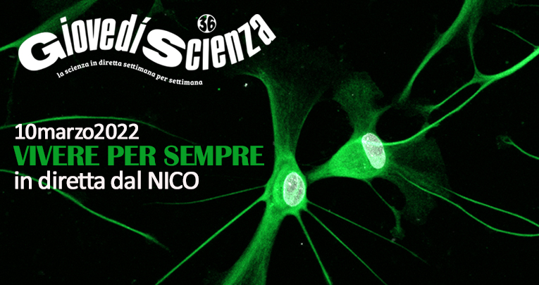
GiovedìScienza racconta la ricerca al NICO
Vivere per sempre.
Una popolazione sempre più longeva, i suoi problemi e le risposte della ricerca
Hai perso la diretta? Guarda ora il video di GiovedìScienza al NICO: una puntata in diretta dai nostri laboratori dedicata alla ricerca sull'invecchiamento.


