Developmental Cell, October 2017
Control of Cell Shape, Neurite Outgrowth, and Migration by a Nogo-A/HSPG Interaction
Anissa Kempf, 1,6,9 Enrica Boda, 2 Jessica C.F. Kwok, 3,8 Rafael Fritz, 4 Valentina Grande, 2 Andrea M. Kaelin, 1 Zorica Ristic, 1 Andre Schmandke, 1 Antonio Schmandke, 1 Bjoern Tews, 5 James W. Fawcett, 3 Olivier Pertz, 4,7 Annalisa Buffo, 2 and Martin E. Schwab 1
Heparan sulfate proteoglycans (HSPGs) critically modulate adhesion-, growth-, and migration-related processes. Here, we show that the transmembrane protein, Nogo-A, inhibits neurite outgrowth and cell spreading in neurons and Nogo-A-responsive cell lines via HSPGs.
The extracellular, active 180 amino acid Nogo-A region, named Nogo-A-Δ20, binds to heparin and brain-derived heparan sulfate glycosaminoglycans (GAGs) but not to the closely related chondroitin sulfate GAGs. HSPGs are required for Nogo-A-Δ20-induced inhibition of adhesion, cell spreading, and neurite outgrowth, as well as for RhoA activation.
Surprisingly, we show that Nogo-A-Δ20 can act via HSPGs independently of its receptor, Sphingosine-1-Phosphate receptor 2 (S1PR2). We thereby identify the HSPG family members syndecan-3 and syndecan-4 as functional receptors for Nogo-A-Δ20. Finally, we show in explant cultures ex vivo that Nogo-A-Δ20 promotes the migration of neuroblasts via HSPGs but not S1PR2.

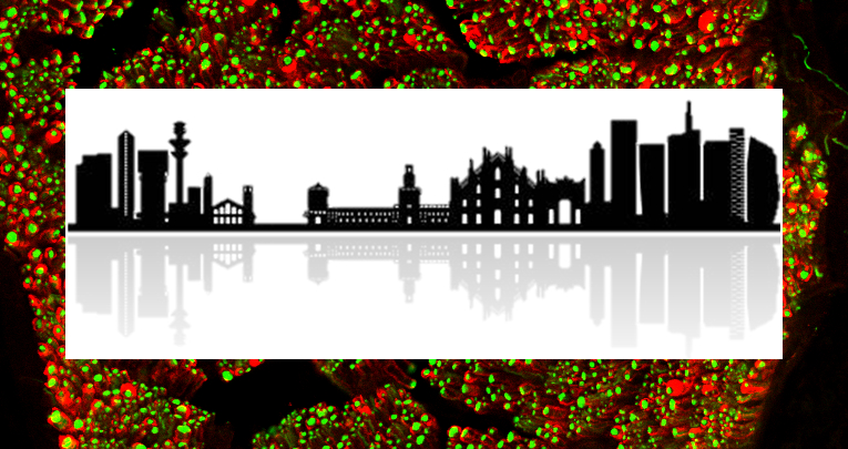
In this picture young neurons (in red) produced by neural stem cells migrate away from the mother cells.
In a first migratory phase, young neurons slide over each other thanks to repulsive signals mediated by the interaction between NoGo-A and Heparan Sulphate Proteoglycans locate on the cells' membrane.

NoGo-A is a membrane protein. Its tail (delta20 segment, in purple) interacts with Heparan Sulphate Proteoglycans (light blue) on the membrane of adjacent cells, thereby inducing diverse effects on brain plasticity.
1
Brain Research Institute, University of Zurich and Department of Health Sciences and Technology, Swiss Federal Institute of Technology (ETH) Zurich, 8057 Zurich, Switzerland
2
Department of Neuroscience, Neuroscience Institute Cavalieri Ottolenghi (NICO), Università degli Studi di Torino, Orbassano, Turin 10043, Italy
3
John van Geest Centre for Brain Repair, Department of Clinical Neurosciences, University of Cambridge, Robinson Way, Cambridge CB2 0PY, UK
4
Institute for Biochemistry and Genetics, Department of Biomedicine, University of Basel, 4058 Basel, Switzerland
5
Schaller Research Group at the University of Heidelberg and the German Cancer Research Center (DKFZ), Molecular Mechanisms of Tumor Invasion, 69120 Heidelberg, Germany
6
Present address: University of Oxford, Centre for Neural Circuits and Behaviour, Oxford OX1 3SR, UK
7
Present address: Institute of Cell Biology, University of Bern, Bern 3012, Switzerland
8
Present address: School of Biomedical Sciences, Faculty of Biological Sciences, University of Leeds, Leeds LS2 9JT, UK
Events & Meetings